
The authors have no financial or proprietary interests related to this paper. In conclusion, although our current findings were based on a single case, additional cases and genetic examination are needed to definitively characterize these rare and unusual associations between congenital grouped pigmentation and albinotic spots in the retina. reported a familial case of a mother and 3 daughters. Similarly, grouped albinotic spots of the retina are also considered to be sporadic, although familial occurrences have rarely been reported. reported a familial case of a mother and daughter who showed an autosomal dominant inheritance, whileDe Jong and Delleman described a father and son with the disease.

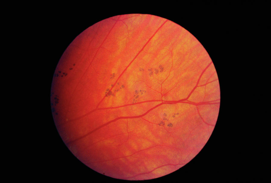
Grouped pigmentation of the retina is considered to be sporadic, although familial occurrences have rarely been reported. To the best of our knowledge, this is the second report of 2 entities, i.e., congenital grouped pigmentation and albinotic spots in the retina, occurring simultaneously in the same individual. Therefore, we considered this condition as congenital grouped pigmentation and albinotic spots in the retina. Their fundus changes were very similar to those observed in the present case. described congenital grouped pigmentation of the retina in a girl and congenital albinotic spots of the retina in her sister. described a case of grouped pigmented lesions with nonpigmented, punctate lesions located within the macula. However, these fundus changes were very similar to those observed in the following 2 reports. In our present case, the size of albinotic spots was smaller than that of the pigmented spots, and the albinotic spots organized in patterns resembling drusen. Congenital grouped albinotic spots are characterized by multiple, variably sized, sharply circumscribed, placoid, white lesions. DiscussionĬongenital grouped pigmentation of the retina is characterized by multiple, grouped, sharply circumscribed, pigmented spots. Right ( a, c) and left ( b, d) fundus photographs of the posterior pole ( a, b) demonstrating albinotic spots, and the peripheral retina ( c, d) demonstrating irregular pigmentation. In addition, a genetic consultation was not performed in this case. Although the patient's mother has undergone a colonoscopy to examine her adenomatous polyposis, abnormal findings were not detected. 3a, b) and an irregular pigmentation in the peripheral retina (fig. Ophthalmoscopic examination of the girl's mother revealed albinotic spots in the posterior pole (fig. 1c, d) and granular hyperfluorescence correlating with the albinotic spots (fig. Fluorescein angiography demonstrated a persistent hypofluorescence correlating with grouped pigmentation of the retina (fig. The albinotic spots were predominantly detected at the posterior pole of both fundi (fig. As a unique co-existing feature, small albinotic spots were identified in the posterior pole (fig. 1a, b), consistent with a grouped pigmentation of the retina or bear track spots. Ophthalmoscopy of both eyes revealed multiple, small, flat, pigmented spots in the peripheral retina (fig. Her visual acuity was 1.2 in both eyes.The anterior segment of both eyes was normal. Her medical and personal histories were unremarkable and her physical examination revealed no abnormalities. Case ReportĪ 10-year-old asymptomatic Japanese girl was referred to our clinic for an ophthalmological examination of bilateral fundus discoloration. Here we present an unusual case of congenital grouped pigmentation of the retina associated with albinotic spots in a 10-year-old Japanese girl.

Congenital albinotic spots of the retina or polar bear tracks are also an uncommon disorder typified by albinotic spots with no visual dysfunction. Detection is usually coincidental during routine ocular examinations. Congenital grouped pigmentation of the retina or bear track spots is an uncommon anomaly characterized by multiple, grouped, sharply circumscribed, pigmented spots with no visual dysfunction.


 0 kommentar(er)
